Anatomy Trains Myofascial Meridians

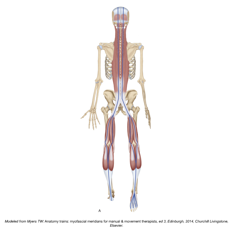
SUPERFICIAL BACK LINE MYOFASCIAL MERIDIAN. PERMISSION JOSEPH E. MUSCOLINO. KINESIOLOGY – THE SKELETAL SYSTEM AND MUSCLE FUNCTION, 3RD ED. (ELSEVIER, 2017).MODELED FROM TOM MYERS’ ANATOMY TRAINS ART.
The Anatomy Trains Myofascial Meridians concept by Tom Myers is quite popular amongst manual and movement therapists. However there has been little scientific evidence to support this concept until recently. Jan Wilke and researchers from Goethe University in Frankfurt, Germany searched for the evidence on the existence of six myofascial meridians, as proposed by Tom Myers in 1997. The study was published in the March 2016 issue of Archives of Physical Medicine and Rehabilitation.
Metastudy
The researchers in this metastudy (a metastudy is a study that reviews a body of previous studies) looked for relevant human anatomical dissection articles published between 1900 and December 2014 in scientific publication databases. The authors appraised peer-reviewed anatomical dissection studies that reported morphological continuity between the muscular components of the myofascial meridians. Also, papers on general anatomy of the corresponding body region were also searched. A continuity between two muscles was only documented if two independent investigators agreed that it was clearly reported. Also, two independent investigators rated methodological quality of included studies. The review identified 6589 articles. Of these, only 62 papers met the inclusion criteria.
Did you know that Digital COMT (Digital Clinical Orthopedic Manual Therapy), Dr. Joe Muscolino’s video streaming subscription service for manual and movement therapists, has six folders with video lessons on Manual Therapy Treatment, including an entire folder on Stretching, as well as a folder on Pathomechanics, another on Anatomy, and many more? Digital COMT adds seven new video lessons each and every week. And nothing ever goes away! Click here for more information.
Results – Strong Evidence Found
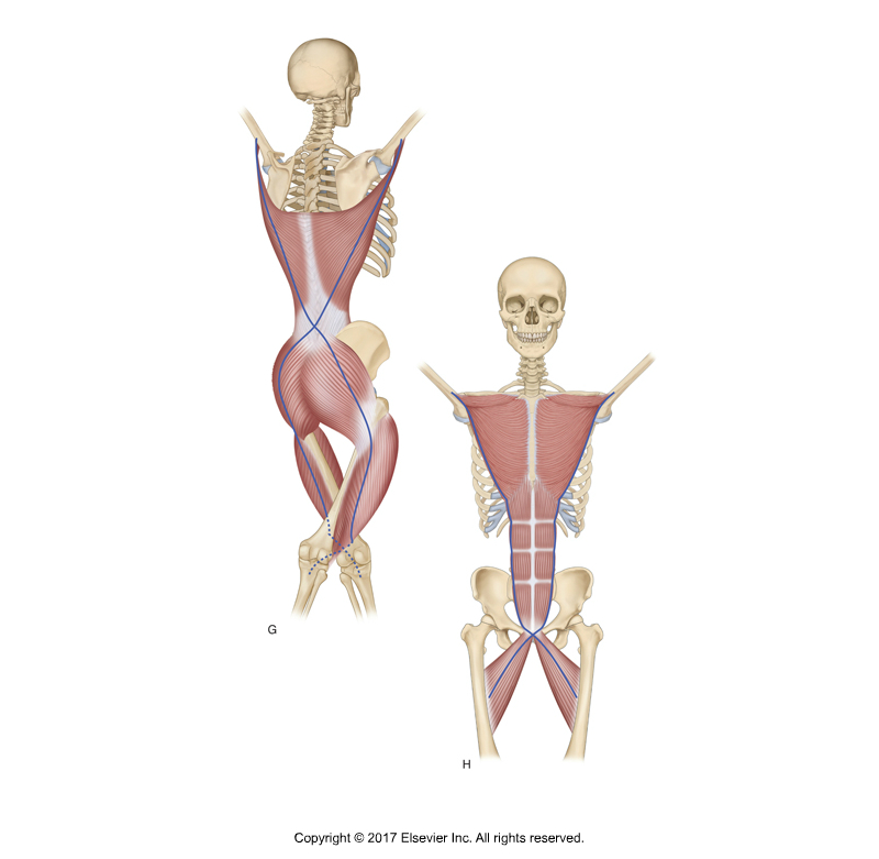
BACK AND FRONT FUNCTIONAL LINE MYOFASCIAL MERIDIANS. PERMISSION JOSEPH E. MUSCOLINO. KINESIOLOGY – THE SKELETAL SYSTEM AND MUSCLE FUNCTION, 3RD ED. (ELSEVIER, 2017). MODELED FROM TOM MYERS’ ANATOMY TRAINS ART.
The review suggests strong evidence for the existence of three myofascial meridians: the Superficial Back Line (all three transitions verified, based on 14 studies), the Back Functional Line (all three transitions verified, eight studies) and the Front Functional Line (both transitions verified, six studies).
Results – Moderate to Strong Evidence Found
Moderate to strong evidence is available for parts of the Spiral Line (five of nine verified transitions, 21 studies) and the Lateral Line (two of five verified transitions, ten studies).
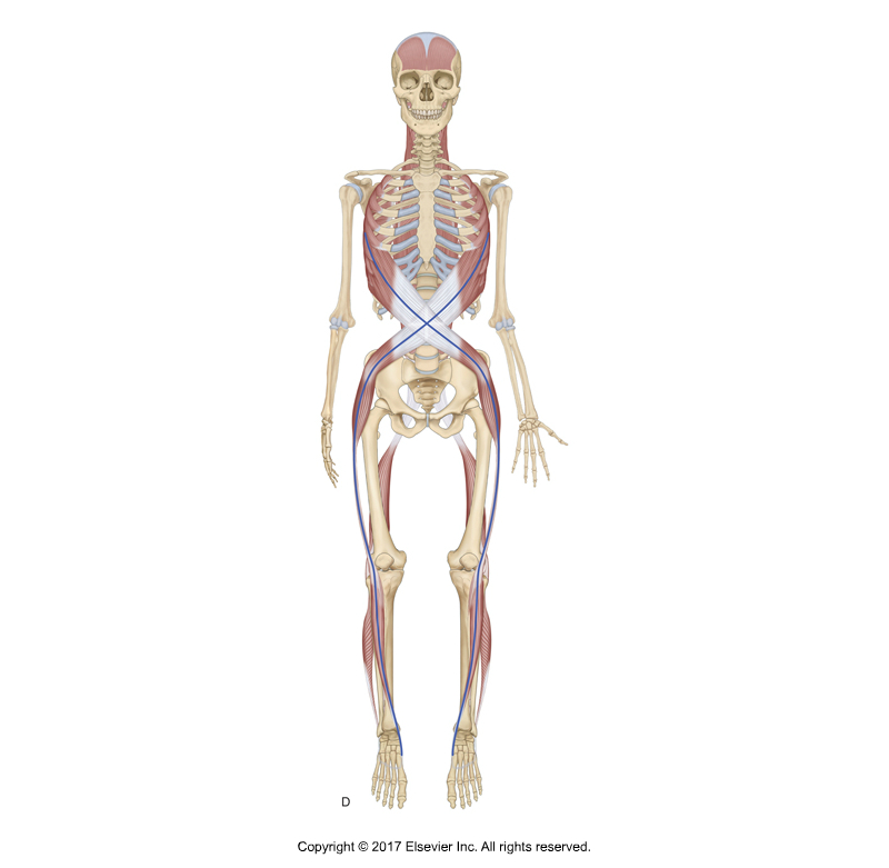
SPIRAL LINE MYOFASCIAL MERIDIAN. PERMISSION JOSEPH MUSCOLINO. KINESIOLOGY – THE SKELETAL SYSTEM AND MUSCLE FUNCTION, 3RD ED. (ELSEVIER, 2017). MODELED FROM ARTWORK IN ANATOMY TRAINS BY TOM MYERS.
For the Spiral Line, continuity between rhomboids and serratus anterior, serratus anterior and external abdominal oblique, and external abdominal oblique and internal abdominal oblique were found. However, no continuity was found below the pelvis.
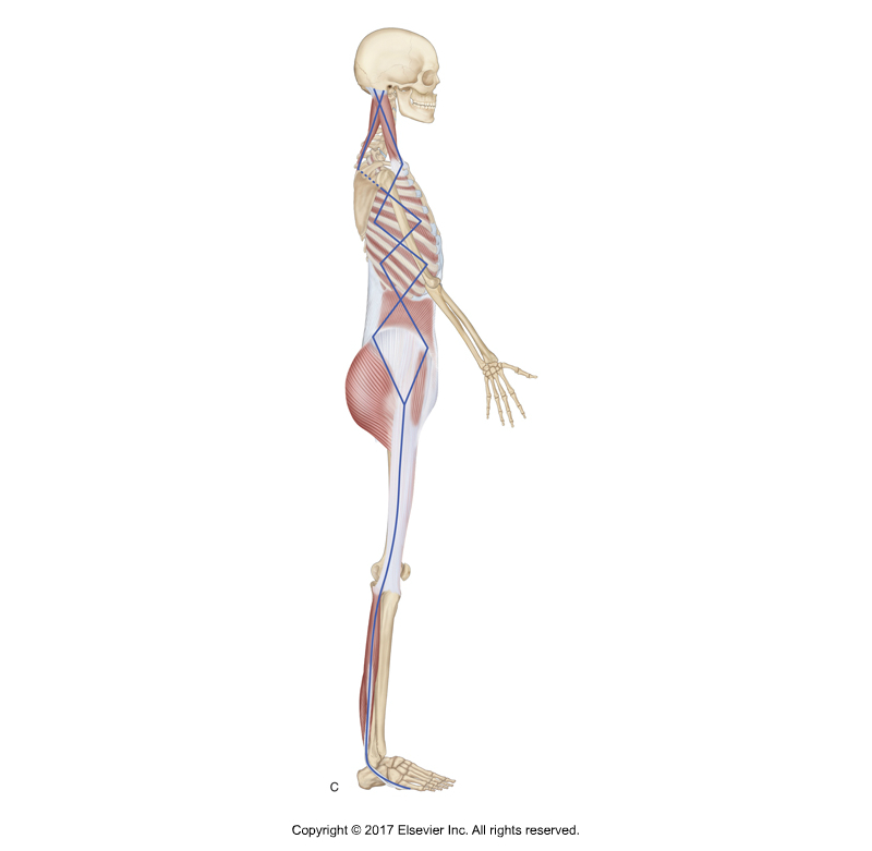
LATERAL LINE MYOFASCIAL MERIDIAN. PERMISSION JOSEPH MUSCOLINO. KINESIOLOGY – THE SKELETAL SYSTEM AND MUSCLE FUNCTION, 3RD ED. (ELSEVIER, 2017). MODELED FROM ARTWORK IN ANATOMY TRAINS BY TOM MYERS.
For the Lateral Line, only continuity iliotibial tract and gluteus maximus/tensor fasciae latae, and gluteus maximus/tensor fasciae latae and abdominal obliques were found.
Results – No Evidence Found
No evidence exists for the Superficial Front Line (no verified transition, seven studies). There is no reported structural connection between the rectus femoris muscle and rectus abdominis. Also, sternalis, which is suggested to be the cranial continuation of rectus abdominis, exists only in a small percentage of the population. Even if present, it does not fuse consistently with the rectus abdominis.
Comment by Joseph Muscolino
I recently published another metastudy review of myofascial meridians. Even though there was certainly some consistency between these two studies, it is interesting to see how one study compared to another can differ. As a general rule, differing results in studies on the same subject can point to not only the inconsistency that can occur from how one study to another is carried out, but also to how a metastudy review can differ from another metastudy review based on how the previous studies are considered (inclusion criteria, etc.). And there is always some subjective judgment both in the conclusion of any one study as well as the manner in which studies are reviewed in a metastudy.
In the particular case of these two studies, they were both done by the same group of researchers. The first study looked at continuity within myofascial meridians while the second study looked at force transmission. What is interesting here is that there can be no force transmission if there is no continuity, right? So evidence of force transmission can be reasonable extrapolated as evidence of continuity (and vice versa).
I will also add something that I added to the previous metastudy blog post article. That is that functional lines myofascial meridians are considered to be ‘functional’ in that their tension transfer is only apparent when the body is functionally in certain positions, which are not likely the positions in which the cadavers are studied (they are more likely to be in position that is close to anatomic position). Certainly, even for this functional continuity within a certain position to be present, there would have to be some continuity in anatomic position as well. However, it is likely that the continuity is much less apparent when the cadaver/body is in anatomic position, and therefore much less likely to be found.
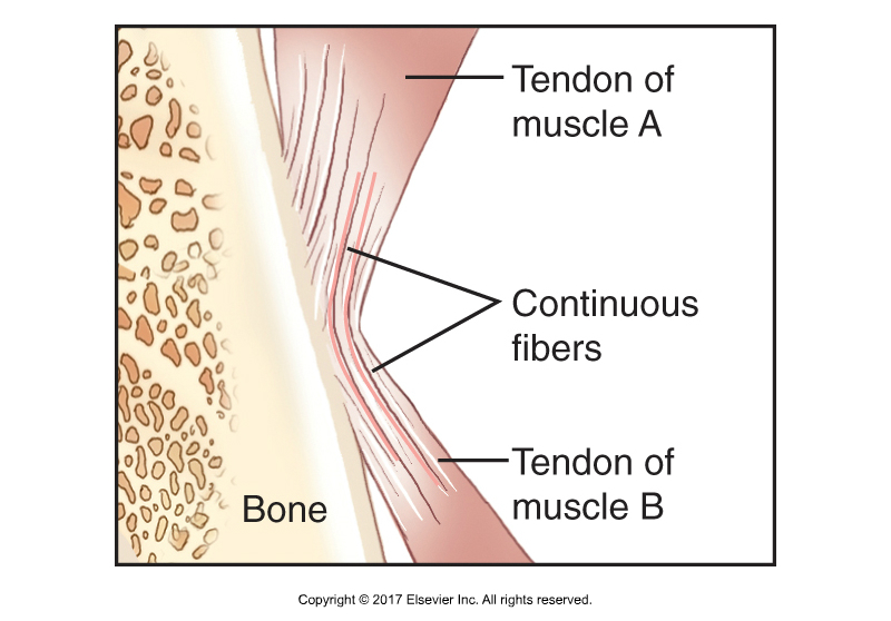
CONCEPT OF A MYOFASCIAL MERIDIAN, ALSO KNOWN AS A MYOFASCIAL CONTINUITY, MYOFASCIAL CHAIN, OR ANATOMY TRAIN. PERMISSION JOSEPH E. MUSCOLINO. KINESIOLOGY – THE SKELETAL SYSTEM AND MUSCLE FUNCTION, 3RD ED. (ELSEVIER, 2017).
Conclusion
The authors suggested that the practical relevance is twofold. First, the existence of the myofascial meridians might help to explain the phenomenon of referred pain. For example, myofascial trigger points in the calf have been shown to elicit pain that radiates to the sole of the foot and the posterior thigh.
A second aspect relates to therapy and training of the musculoskeletal system. Treatment according to myofascial meridians could be effective in reducing back pain. Several studies have shown that low back pain patients display reduced hamstring flexibility. Overload injuries in competitive sports represent another entity of pathologies, which possibly occur due to the presence of myofascial meridians. Recent studies indicate that tightness of the gastrocnemius and the hamstrings are associated with plantar fasciitis. Groin pain or athletic pubalgia is suggested to be provoked by a tight adductor longus and a weak rectus abdominis. Strain transmission along meridians would both open a new frontier for the understanding of referred pain and provide a rationale for the development of more body-wide holistic treatment approaches.
This blog post article was created in collaboration with www.terrarosa.com.au.
(Click here for the blog post article Increased Muscle Activation in Back Line Myofascial Continuity.)
Did you know that Digital COMT (Digital Clinical Orthopedic Manual Therapy), Dr. Joe Muscolino’s video streaming subscription service for manual and movement therapists, has six folders with video lessons on Manual Therapy Treatment, including an entire folder on Stretching, as well as a folder on Pathomechanics, another on Anatomy, and many more? Digital COMT adds seven new video lessons each and every week. And nothing ever goes away! Click here for more information.